Abstract
Introduction
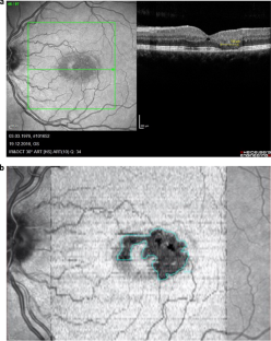
Macular Telangiectasia type 2 (MacTel) is a bilateral neurodegenerative disease associated with dysfunction in the serine and lipid metabolism resulting in loss of Muller cells and photoreceptors. Typical structural changes include vascular abnormalities, loss of retinal transparency, redistribution of macular pigment and thinning of the central retina with photoreceptor loss. The presence and extent of photoreceptor loss, as visible on Optical Coherence Tomography (OCT) (“disease severity scale”), correlate with functional loss and the limitation of photoreceptor loss appears to be the most promising therapeutic approach. Ongoing clinical trials of ciliary neurotrophic factor (CNTF) implants for the treatment of MacTel are using this outcome to evaluate efficacy. An ideal outcome measure provides the ability to quantify the extent of the disease progression with precision and reproducibility.
Methods
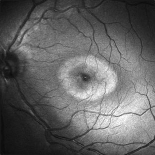
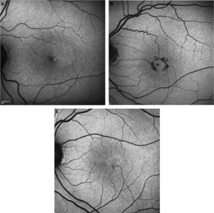
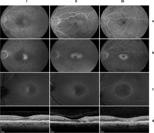
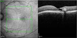
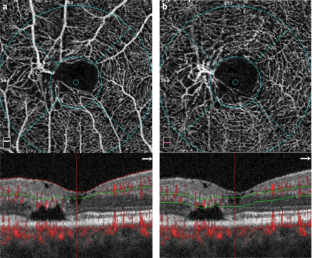
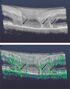
This review describes the changes and findings on different imaging techniques including fluorescein- and OCT angiography, blue light reflectance, 1- and 2-wavelength autofluorescence and OCT.
Results
The possibilities of objective quantification of the severity of MacTel and correlation with functional characteristics such as best-corrected visual acuity (BCVA) and microperimetry and their applications as quantitative imaging endpoints for clinical treatment trials are discussed. OCT and especially en face OCT could be demonstrated as precise and reproducible methods to quantify the area of photoreceptor loss, which correlated highly significantly with functional loss in microperimetry.
Conclusion
The analysis of the area of photoreceptor loss on en face OCT is the most reliable imaging endpoint for treatment trials in MacTel. This method is already being used in ongoing randomized trials.
Access options
Subscribe to Journal
Get full journal access for 1 year
259,00 €
only 21,58 € per issue
Tax calculation will be finalised during checkout.
Rent or Buy article
Get time limited or full article access on ReadCube.
from$8.99
All prices are NET prices.
References
- 1.
Charbel Issa P, Gillies MC, Chew EY, Bird AC, Heeren TF, Peto T, et al. Macular telangiectasia type 2. Prog Retin Eye Res. 2013;34:49–77.
- 2.
Powner MB, Gillies MC, Tretiach M, Scott A, Guymer RH, Hageman GS, et al. Perifoveal Müller cell depletion in a case of macular telangiectasia type 2. Ophthalmology. 2010;117:2407–16.
- 3.
Powner MB, Gillies MC, Zhu M, Vevis K, Hunyor AP, Fruttiger M. Loss of Müller’s cells and photoreceptors in macular telangiectasia type 2. Ophthalmology. 2013;120:2344–52.
- 4.
Gantner ML, Eade K, Wallace M, Handzlik MK, Fallon R, Trombley J, et al. Serine and lipid metabolism in macular disease and peripheral neuropathy. N Engl J Med. 2019;381:1422–33.
- 5.
Gass JD, Oyakawa RT. Idiopathic juxtafoveolar retinal telangiectasis. Arch Ophthalmol. 1982;100:769–80.
- 6.
Gass JDM, Blodi BA. Idiopathic juxtafoveolar retinal telangiectasis: update of classification and follow-up study. Ophthalmology. 1993;100:1536–46.
- 7.
Esposti SD, Egan C, Bunce C, Moreland JD, Bird AC, Robson RG. Macular pigment parameters in patients with macular telangiectasia (MacTel) and normal subjects: implications of a novel analysis. Investig Ophthalmol Vis Sci. 2012;53:6568–75.
- 8.
Zeimer MB, Padge B, Heimes B, Pauleikhoff D. Idiopathic macular telangiectasia type 2: distribution of macular pigment and functional investigations. Retina. 2010;30:586–95.
- 9.
Charbel Issa P, Berendschot TT, Staurenghi G, Holz FG, Scholl HP. Confocal blue reflectance imaging in type 2 idiopathic macular telangiectasia. Investig Ophthalmol Vis Sci. 2008;49:1172–7.
- 10.
Sallo FB, Leung I, Zeimer M, Clemons TE, Dubis AM, Fruttiger M, et al. Abnormal retinal reflectivity to short-wavelength light in type 2 idiopathic macular teleangiectasis. Retina. 2018;38:S79–88.
- 11.
Tzaridis S, Heeren TFC, Mai C, Thiele S, Holz FG, Charbel Issa P, et al. Right-angled vessels in macular telangiectasia type 2. Br J Ophthalmol. 2019. https://doi.org/10.1136/bjophthalmol-2018-313364. Online ahead of print.
- 12.
Gaudric A, Ducos de Lahitte G, Cohen SY, Massin P, Haouchine B. Optical coherence tomography in group 2A idiopathic juxtafoveolar retinal telangiectasis. Arch Ophthalmol. 2006;124:1410–9.
- 13.
Sallo FB, Peto T, Egan C, Wolf-Schnurrbusch UE, Clemons TE, Gillies MC, et al. The IS/OS junction layer in the natural history of type 2 idiopathic macular telangiectasia. Investig Ophthalmol Vis Sci. 2012;53:7889–95.
- 14.
Sallo FB, Leung I, Clemons TE, Peto T, Bird AC, Pauleikhoff D. Multimodal imaging in type 2 idiopathic macular teleangiectasis. Retina. 2015;35:742–9.
- 15.
Mukherjee D, Lad EM, Vann RR, Jaffe SJ, Clemons TE, Friedlander M, et al. Correlation between macular integrity assessment and optical coherence tomography imaging of ellipsoid zone in macular telangiectasia type 2. Investig Ophthalmol Vis Sci. 2017;58:291–9.
- 16.
Heeren TFC, Kitka D, Florea D, Clemons TE, Chew EY, Bird AC, et al. Longitudinal correlation of ellipsoid zone loss and functional loss in Macular Telangiectasia Type 2. Retina. 2018;38:S20–26.
- 17.
Okada M, Robson AG, Egan CA, Sallo FB, Esposti SD, Heeren TFC, et al. Electrophysiological characterization of Macular Telangiectasia Type 2 and structure-function correlation. Retina. 2018;38:S33–42.
- 18.
Peto T, Heeren TFC, Clemons TE, Sallo FB, Leung I, Chew EY, et al. Correlation of clinical and functional progression with visual acuity loss in Macular Telangiectasia Type 2. MacTel Project Report No. 6–The MacTel Research Group. Retina. 2018;38:S8–13.
- 19.
Pauleikhoff D, Bonelli R, Dubis AM, Gunnemann F, Rothaus K, Charbel Issa P, et al. Progression characteristics of ellipsoid zone loss in macular teleangiectasia type 2. Acta Ophthalmol. 2019;97:998–1005.
- 20.
Mueller S, Gunnemann F, Rothaus K, Book M, Faatz H, Bird AC, et al. Incidence and phenotypical variation of outer retina-associated hyperreflectivity in macular telangiectasia type 2. Br J Ophthalmol. 2021;105:573–6.
- 21.
Gaudric A, Krivosic V, Tadayoni R. Outer retina capillary invasion and ellipsoid zone loss in Macular Teleangiectasia Type 2 imaged by OCT angiography. Retina. 2015;35:2300–6.
- 22.
Spaide RF, Yannuzzi LA, Maloca PM. Retinal-choroidal anastomosis in macular teleangictasis type 2. Retina. 2018;38:1920–9.
- 23.
Breazzano MP, Yannuzzi LA, Spaide RF. Characterizing retinal-choroidal anastomosis in macular teleangiectasia type 2 with optical coherence tomography angiography. Retina. 2020;40:92–8.
- 24.
Pauleikhoff D, Gunnemann F, Book M, Rothaus K. Progression of vascular changes in macular telangiectasia type 2: comparison between SD-OCT and OCT angiography. Graefes Arch Clin Exp Ophthalmol. 2019;257:1381–92.
- 25.
Tzaridis S, Hess K, Friedlander M, Holz FG. Optical coherence tomography-angiography for monitoring neovascularisations in macular telangiectasia type 2. Br J Ophthalmol. 2021;105:735–40.
- 26.
Heeren TFC, Chew EY, Clemons T, Fruttiger M, Balaskas K, Schwartz R, et al. MacTel Study Group. Macular telangiectasia type 2 - visual acuity, disease endstage and the MacTel area. MacTel Project Report No. 8. Ophthalmology. 2020;127:1539–48.
- 27.
Charbel Issa P, Heeren TF, Kupitz EH, Holz FG, Berendschot TT. Very early disease manifestations of macular telangiectasia type 2. Retina. 2016;36:524–34.
- 28.
Heeren TF, Clemons T, Scholl HP, Bird AC, Holz FG, Charbel Issa P. Progression of vision loss in macular telangiectasia type 2. Investig Ophthalmol Vis Sci. 2015;56:3905–12.
- 29.
Charbel Issa P, Finger RP, Helb HM, Holz FG, Scholl HP. A new diagnostic approach in patients with type 2 macular telangiectasia: confocal reflectance imaging. Acta Ophthalmol. 2008;86:464–5.
- 30.
Bottoni F, Eandi CM, Pedenovi S, Staurenghi G. Integrated clinical evaluation of Type 2A idiopathic juxtafoveolar retinal telangiectasis. Retina. 2010;30:317–26.
- 31.
Jindal A, Choudhury H, Pathengay A, Flynn HW Jr. A novel clinical sign in macular telangiectasia type 2. Ophthalmic Surg Lasers Imaging Retin. 2015;46:134–6.
- 32.
Okada M, Heeren TFC, Egan CA, Rocco V, Bonelli R, Fruttiger M. Effect of dark adaptation and bleaching on Blue Light Reflectance imaging in Macular Teleangiectasia Type 2. Retina. 2018;38:89–96.
- 33.
Pauleikhoff L, Heeren TFC, Gliem M, Lim E, Pauleikhoff D, Holz FG, et al. Fundus autofluorescence imaging in macular telangiectasia type 2 - MacTel Study Report Number 9: fundus autofluorescence in macular telangiectasia type 2. Am J Ophthalmol. 2021;228:27–34.
- 34.
Zeimer MB, Krömer I, Spital G, Lommatzsch A, Pauleikhoff D. Macular telangiectasia: patterns of distribution of macular pigment and response to supplementation. Retina. 2010;3:1282–93.
- 35.
Zeimer MB, Spital G, Heimes B, Lommatzsch A, Pauleikhoff D. Macular telangiectasia-changes in macular pigment optical density during a 5-year follow-up. Retina. 2014;34:920–8.
- 36.
Zeimer MB, Sallo FB, Spital G, Heimes B, Lommatzsch A, Pauleikhoff D. Correlation of optical coherence tomography and macular pigment optical density measurements in type 2 macular teleangiectasia. Retina. 2016;36:535–44.
- 37.
Theelen T, Berendschot TT, Boon CJ, Hoyng CB, Klevering BJ. Analysis of visual pigment by fundus autofluorescence. Exp Eye Res. 2008;8:296–304.
- 38.
Trieschmann M, Heimes B, Hense HW, Pauleikhoff D. Macular pigment optical density measurement in autofluorescence imaging: comparison of one- and two-wavelength methods. Graefes Arch Clin Exp Ophthalmol. 2006;244:1565–74.
- 39.
Helb HM, Charbel Issa P, RL VDV, Berendschot TT, Scholl HP, Holz FG. Abnormal macular pigment distribution in type 2 idiopathic macular telangiectasia. Retina. 2008;28:808–16.
- 40.
Charbel Issa P, van der Veen RL, Stijfs A, Holz FG, Scholl HP, Berendschot TT. Quantification of reduced macular pigment optical density in the central retina in macular telangiectasia type 2. Exp Eye Res. 2009;89:25–31.
- 41.
Sallo FB, Peto T, Egan C, Wolf-Schnurrbusch UE, Clemons TE, Gillies MC, et al. “En face” OCT imaging of the IS/OS junction line in type 2 idiopathic macular telangiectasia. Investig Ophthalmol Vis Sci. 2012;5:6145–52.
- 42.
Krivosic V, Lavia C, Aubineau A, Tadayoni R, Alain Gaudric A. OCT of outer retinal hyperreflectivity, neovascularization, and pigment in macular telangiectasia type 2. Ophthalmol Retina. 2020;19:2468–6530.
- 43.
Tzaridis S, Friedlander M, Macular Telangiectasia Type 2-Phase 2 CNTF Research Group. Functional relevance of hyper-reflectivity in macular telangiectasia type 2. Investig Ophthalmol Vis Sci. 2021;62:6. https://doi.org/10.1167/iovs.62.3.6.
- 44.
Breazzano MP, Yannuzzi LA, Spaide RF. Genesis of retinal-choroidal anastomosis in macular telangiectasia type 2: a longitudinal analysis. Retina. 2020;40:92–8.
- 45.
Chew EY, Clemons TE, Peto T, Sallo FB, Ingerman A, Tao W, et al. Ciliary neurotrophic factor for macular telangiectasia type 2: results from a phase 1 safety trial. Am J Ophthalmol. 2015;159:659–66.
- 46.
Chew EY, Clemons TE, Jaffe GJ, Johnson CA, Farsiu S, Lad EM, et al. Effect of ciliary neurotrophic factor on retinal neurodegeneration in patients with macular telangiectasia type 2: a randomized clinical trial. Ophthalmology. 2019;126:540–9.
- 47.
Jia Y, Tan O, Tokayer J, Potsaid B, Wang Y, Liu JJ, et al. Split-spectrum amplitude-decorrelation angiography with optical coherence tomography. Opt Express. 2012;20:4710–25.
- 48.
Zeimer M, Gutfleisch M, Heimes B, Spital G, Lommatzsch A, Pauleikhoff D. Association between changes in macular vasculature in optical coherence tomography- and fluorescein-angiography and distribution of macular pigment in type 2 idiopathic macular teleangiectasia. Retina. 2015;35:2307–16.
- 49.
Faatz H, Rothaus K, Gunnemann F, Gutfleisch M, Heimes-Bussmann B, Lommatzsch A, et al. Changes in OCT angiography of Type 2 CNV in neovascular AMD during anti-VEGF treatment. Klin Monatsbl Augenheilkd. 2017;234:1125–31.
- 50.
Faatz H, Farecki ML, Rothaus, Gunnemann F, Gutfleisch M, Lommatzsch A. Optical coherence tomography angiography of type 1 and 2 neovascularisations in age-related macular degeneration under anti-VEGF therapy: Evaluation of a new quantitative method. Eye. 2019;33:1466–71.
- 51.
Zhang Q, Wang RK, Chen CL, Legarreta AD, Durbin MK, An L, et al. Swept source optical coherence tomography angiography of neovascular macular teleangiectasia type 2. Retina. 2015;35:2285–99.
- 52.
Runkle AP, Kaiser PK, Srivastava SK, Schachat AP, Reese JL, Ehlers JP. OCT angiography and ellipsoid zone mapping of macular telangiectasia type 2 from the AVATAR study. Investig Ophthalmol Vis Sci. 2017;58:3683–9.
- 53.
Leung I, Sallo FB, Bonelli R, Clemons TE, Pauleikhoff D, Chew EY, et al. Characteristics of pigmented lesions in type 2 idiopathic macular teleangiectasia. Retina. 2018;38:43–50.
- 54.
Ray TA, Cochran KJ, Kay JN. The enigma of CRB1 and CRB1 retinopathies. Adv Exp Med Biol. 2019;1185:251–5.
- 55.
Usui-Ouchi A, Usui Y, Kurihara T, Aguilar E, Dorrell MI, Ideguchi Y, et al. Retinal microglia are critical for subretinal neovascular formation. JCI Insight. 2020;5:e137317.
- 56.
Spaide RF, Jaffe GJ, Sarraf D, Freund KB, Sadda SR, Staurenghi G, et al. Consensus nomenclature for reporting neovascular age-related macular degeneration data: consensus on neovascular age-related macular degeneration nomenclature study group. Ophthalmology. 2020;127:616–36.
- 57.
Kupitz EH, Heeren TFC, Holz FG, Charbel Issa P. Poor long-term outcome of Anti-VEGF-Therapy in nonproliferative Macular Teleangiectasia Type 2. Retina. 2015;35:2619–26.
- 58.
Tapia Quijada HE, Mantolan Sarmiento C, Gómez Perera S, Serrano García M, Betancor Caro N, Haro Álvarez B. OCT-angiography for diagnosis and response to treatment of subretinal neovascularization secondary to idiopathic macular telangiectasia type 2. Arch Soc Esp Oftalmol. 2021. https://doi.org/10.1016/j.oftal.2020.11.020. Online ahead of print.
Author information
Affiliations
Contributions
DP: structuring and editing of the manuscript. LP, EYC: editing of the manuscript.
Corresponding author
Ethics declarations
Competing interests
The authors declare no competing interests.
Additional information
Publisher’s note Springer Nature remains neutral with regard to jurisdictional claims in published maps and institutional affiliations.
Rights and permissions
About this article
Cite this article
Pauleikhoff, D., Pauleikhoff, L. & Chew, E.Y. Imaging endpoints for clinical trials in MacTel type 2. Eye (2021). https://ift.tt/3iKzLxl
-
Received:
-
Revised:
-
Accepted:
-
Published:
"type" - Google News
August 13, 2021 at 10:43PM
https://ift.tt/3sgl5c9
Imaging endpoints for clinical trials in MacTel type 2 | Eye - Nature.com
"type" - Google News
https://ift.tt/2WhN8Zg
https://ift.tt/2YrjQdq
Bagikan Berita Ini


0 Response to "Imaging endpoints for clinical trials in MacTel type 2 | Eye - Nature.com"
Post a Comment