Abstract
Type 2 diabetes mellitus (T2D), a major cause of worldwide morbidity and mortality, is characterized by dysfunction of insulin-producing pancreatic islet β cells1,2. T2D genome-wide association studies (GWAS) have identified hundreds of signals in non-coding and β cell regulatory genomic regions, but deciphering their biological mechanisms remains challenging3,4,5. Here, to identify early disease-driving events, we performed traditional and multiplexed pancreatic tissue imaging, sorted-islet cell transcriptomics and islet functional analysis of early-stage T2D and control donors. By integrating diverse modalities, we show that early-stage T2D is characterized by β cell-intrinsic defects that can be proportioned into gene regulatory modules with enrichment in signals of genetic risk. After identifying the β cell hub gene and transcription factor RFX6 within one such module, we demonstrated multiple layers of genetic risk that converge on an RFX6-mediated network to reduce insulin secretion by β cells. RFX6 perturbation in primary human islet cells alters β cell chromatin architecture at regions enriched for T2D GWAS signals, and population-scale genetic analyses causally link genetically predicted reduced RFX6 expression with increased T2D risk. Understanding the molecular mechanisms of complex, systemic diseases necessitates integration of signals from multiple molecules, cells, organs and individuals, and thus we anticipate that this approach will be a useful template to identify and validate key regulatory networks and master hub genes for other diseases or traits using GWAS data.
This is a preview of subscription content, access via your institution
Access options
Access Nature and 54 other Nature Portfolio journals
Get Nature+, our best-value online-access subscription
$29.99 / 30 days
cancel any time
Subscribe to this journal
Receive 51 print issues and online access
$199.00 per year
only $3.90 per issue
Rent or buy this article
Prices vary by article type
from$1.95
to$39.95
Prices may be subject to local taxes which are calculated during checkout


Data availability
Raw genotyping, bulk RNA-seq and single-nucleus multiome data are available via the European Genome-Phenome Archive (EGA; RRID SCR_004944) as study EGAS00001006273. Raw imaging datasets are available from the authors upon request. Processed imaging data is available via Pancreatlas (RRID SCR_018567; https://pancreatlas.org) and at https://zenodo.org/doi/10.5281/zenodo.8125025; weighted gene co-expression network analysis is available at https://theparkerlab.shinyapps.io/Islet-RNAseq-WGCNA/; and processed pseudoislet multiome data are available at https://theparkerlab.med.umich.edu/data/public/cellbrowser/?ds=Pseudoislet10XMultiome. In addition, intermediate processed results (expression matrices and others) are available via Zenodo (https://zenodo.org/doi/10.5281/zenodo.6515986). All datasets are linked from http://theparkerlab.org/manuscripts/2021_islet-rfx6/. Source data are provided with this paper.
Code availability
Packages and algorithms used for transcriptomic and cell neighbourhood analyses are provided in the Github repositories https://github.com/ParkerLab and https://github.com/liu-bioinfo-lab.
References
-
Kahn, S. E., Hull, R. L. & Utzschneider, K. M. Mechanisms linking obesity to insulin resistance and type 2 diabetes. Nature 444, 840–846 (2006).
-
Halban, P. A. et al. β-cell failure in type 2 diabetes: postulated mechanisms and prospects for prevention and treatment. Diabetes Care 37, 1751–1758 (2014).
-
Mahajan, A. et al. Fine-mapping type 2 diabetes loci to single-variant resolution using high-density imputation and islet-specific epigenome maps. Nat. Genet. 50, 1505–1513 (2018).
-
Rai, V. et al. Single-cell ATAC-seq in human pancreatic islets and deep learning upscaling of rare cells reveals cell-specific type 2 diabetes regulatory signatures. Mol. Metab. 32, 109–121 (2019).
-
Chiou, J. et al. Single-cell chromatin accessibility identifies pancreatic islet cell type- and state-specific regulatory programs of diabetes risk. Nat. Genet. 53, 455–466 (2021).
-
Ahlqvist, E., Prasad, R. B. & Groop, L. Subtypes of type 2 diabetes determined from clinical parameters. Diabetes 69, 2086–2093 (2020).
-
Redondo, M. J. et al. The clinical consequences of heterogeneity within and between different diabetes types. Diabetologia 63, 2040–2048 (2020).
-
Weitz, J., Menegaz, D. & Caicedo, A. Deciphering the complex communication networks that orchestrate pancreatic islet function. Diabetes 70, 17–26 (2020).
-
Vujkovic, M. et al. Discovery of 318 new risk loci for type 2 diabetes and related vascular outcomes among 1.4 million participants in a multi-ancestry meta-analysis. Nat. Genet. 52, 680–691 (2020).
-
Mahajan, A. et al. Multi-ancestry genetic study of type 2 diabetes highlights the power of diverse populations for discovery and translation. Nat. Genet. 54, 560–572 (2022).
-
Parker, S. C. J. et al. Chromatin stretch enhancer states drive cell-specific gene regulation and harbor human disease risk variants. Proc. Natl Acad. Sci. USA 110, 17921–17926 (2013).
-
Trynka, G. et al. Chromatin marks identify critical cell types for fine mapping complex trait variants. Nat. Genet. 45, 124–130 (2013).
-
Pasquali, L. et al. Pancreatic islet enhancer clusters enriched in type 2 diabetes risk-associated variants. Nat. Genet. 46, 136–43 (2014).
-
Walker, J. T., Saunders, D. C., Brissova, M. & Powers, A. C. The human islet: mini-organ with mega-impact. Endocr. Rev. 42, bnab010 (2021).
-
Brissova, M. et al. Assessment of human pancreatic islet architecture and composition by laser scanning confocal microscopy. J. Histochem. Cytochem. 53, 1087–1097 (2005).
-
Dai, C. et al. Stress-impaired transcription factor expression and insulin secretion in transplanted human islets. J. Clin. Invest. 126, 1857–1870 (2016).
-
Wigger, L. et al. Multi-omics profiling of living human pancreatic islet donors reveals heterogeneous beta cell trajectories towards type 2 diabetes. Nat. Metab. 3, 1017–1031 (2021).
-
Camunas-Soler, J. et al. Patch-seq links single-cell transcriptomes to human islet dysfunction in diabetes. Cell Metab. 31, 1017–1031.e4 (2020).
-
Shapira, S. N., Naji, A., Atkinson, M. A., Powers, A. C. & Kaestner, K. H. Understanding islet dysfunction in type 2 diabetes through multidimensional pancreatic phenotyping: The Human Pancreas Analysis Program. Cell Metab. 34, 1906–1913 (2022).
-
Albrechtsen, N. J. W. et al. The liver–α-cell axis and type 2 diabetes. Endocr. Rev. 40, 1353–1366 (2019).
-
Wu, M. et al. Single-cell analysis of the human pancreas in type 2 diabetes using multi-spectral imaging mass cytometry. Cell Rep. 37, 109919 (2021).
-
Dam, T. J. Pvan et al. CiliaCarta: an integrated and validated compendium of ciliary genes. PLoS ONE 14, e0216705 (2019).
-
Smith, S. B. et al. Rfx6 directs islet formation and insulin production in mice and humans. Nature 463, 775–780 (2010).
-
Patel, K. A. et al. Heterozygous RFX6 protein truncating variants are associated with MODY with reduced penetrance. Nat. Commun. 8, 888 (2017).
-
Varshney, A. et al. Genetic regulatory signatures underlying islet gene expression and type 2 diabetes. Proc. Natl Acad. Sci. USA 114, 2301–2306 (2017).
-
Walker, J. T. et al. Integrated human pseudoislet system and microfluidic platform demonstrates differences in G-protein-coupled-receptor signaling in islet cells. JCI Insight 5, e137017 (2020).
-
Viñuela, A. et al. Genetic variant effects on gene expression in human pancreatic islets and their implications for T2D. Nat. Commun. 11, 4912 (2020).
-
Kahn, S. E., Zraika, S., Utzschneider, K. M. & Hull, R. L. The beta cell lesion in type 2 diabetes: there has to be a primary functional abnormality. Diabetologia 52, 1003–1012 (2009).
-
Meier, J. J. & Bonadonna, R. C. Role of reduced β-cell mass versus impaired β-cell function in the pathogenesis of type 2 diabetes. Diabetes Care 36, S113–S119 (2013).
-
Cohrs, C. M. et al. Dysfunction of persisting β cells is a key feature of early type 2 diabetes pathogenesis. Cell Rep. 31, 107469 (2020).
-
McCarthy, M. I. Painting a new picture of personalised medicine for diabetes. Diabetologia 60, 793–799 (2017).
-
Chandra, V. et al. RFX6 regulates insulin secretion by modulating Ca2+ homeostasis in human β cells. Cell Rep. 9, 2206–2218 (2014).
-
Piccand, J. et al. Rfx6 maintains the functional identity of adult pancreatic β cells. Cell Rep. 9, 2219–2232 (2014).
-
Choksi, S. P., Lauter, G., Swoboda, P. & Roy, S. Switching on cilia: transcriptional networks regulating ciliogenesis. Development 141, 1427–1441 (2014).
-
Piasecki, B. P., Burghoorn, J. & Swoboda, P. Regulatory factor X (RFX)-mediated transcriptional rewiring of ciliary genes in animals. Proc. Natl Acad. Sci. USA 107, 12969–12974 (2010).
-
Kurki, M. I. et al. FinnGen provides genetic insights from a well-phenotyped isolated population. Nature 613, 508–518 (2023).
-
Iotchkova, V. et al. GARFIELD classifies disease-relevant genomic features through integration of functional annotations with association signals. Nat. Genet. 51, 343–353 (2019).
-
Gloyn, A. L. et al. Every islet matters: improving the impact of human islet research. Nat. Metab. 4, 970–977 (2022).
-
Balamurugan, A. N., Chang, Y., Fung, J. J., Trucco, M. & Bottino, R. Flexible management of enzymatic digestion improves human islet isolation outcome from sub‐optimal donor pancreata. Am. J. Transplant. 3, 1135–1142 (2003).
-
Dai, C. et al. Age-dependent human β cell proliferation induced by glucagon-like peptide 1 and calcineurin signaling. J. Clin. Invest. 127, 3835–3844 (2017).
-
Brissova, M. et al. α cell function and gene expression are compromised in type 1 diabetes. Cell Rep. 22, 2667–2676 (2018).
-
Brissova, M. et al. Islet microenvironment, modulated by vascular endothelial growth factor-A signaling, promotes β cell regeneration. Cell Metab. 19, 498–511 (2014).
-
Brissova, M. et al. The Integrated Islet Distribution Program answers the call for improved human islet phenotyping and reporting of human islet characteristics in research articles. Diabetologia 62, 1312–1314 (2019).
-
Kayton, N. S. et al. Human islet preparations distributed for research exhibit a variety of insulin-secretory profiles. Am. J. Physiol. Endocrinol. Metab. 308, E592–E602 (2015).
-
Fitzmaurice, G. M., Laird, N. M. & Ware, J. H. Applied Longitudinal Analysis (Wiley, 2011).
-
Shultz, L. D. et al. Human lymphoid and myeloid cell development in NOD/LtSz-scid IL2Rγnull mice engrafted with mobilized human hemopoietic stem cells. J. Immunol. 174, 6477–6489 (2005).
-
Dai, C. et al. Tacrolimus- and sirolimus-induced human β cell dysfunction is reversible and preventable. JCI Insight 5, e130770 (2020).
-
Dorrell, C. et al. Transcriptomes of the major human pancreatic cell types. Diabetologia 54, 2832 (2011).
-
Saunders, D. C. et al. Ectonucleoside triphosphate diphosphohydrolase-3 antibody targets adult human pancreatic β cells for in vitro and in vivo analysis. Cell Metab. 29, 745–754.e4 (2019).
-
Dorrell, C. et al. Human islets contain four distinct subtypes of β cells. Nat. Commun. 7, 11756 (2016).
-
Haliyur, R. et al. Human islets expressing HNF1A variant have defective β cell transcriptional regulatory networks. J. Clin. Invest. 129, 246–251 (2018).
-
Marzban, L., Park, K. & Verchere, C. B. Islet amyloid polypeptide and type 2 diabetes. Exp. Gerontol. 38, 347–351 (2003).
-
Westermark, P., Andersson, A. & Westermark, G. T. Islet amyloid polypeptide, islet amyloid, and diabetes mellitus. Physiol. Rev. 91, 795–826 (2011).
-
Hart, N. J. et al. Cystic fibrosis–related diabetes is caused by islet loss and inflammation. JCI Insight 3, e98240 (2018).
-
Noguchi, G. M. & Huising, M. O. Integrating the inputs that shape pancreatic islet hormone release. Nat. Metab. 1, 1189–1201 (2019).
-
Black, S. et al. CODEX multiplexed tissue imaging with DNA-conjugated antibodies. Nat. Protoc. 16, 3802–3835 (2021).
-
Blondel, V. D., Guillaume, J.-L., Lambiotte, R. & Lefebvre, E. Fast unfolding of communities in large networks. J. Stat. Mech. Theory Exp. 2008, P10008 (2008).
-
Luhn, H. P. The automatic creation of literature abstracts. IBM J. Res. Dev. 2, 159–165 (1958).
-
Schürch, C. M. et al. Coordinated cellular neighborhoods orchestrate antitumoral immunity at the colorectal cancer invasive front. Cell 182, 1341–1359.e19 (2020).
-
Dobin, A. et al. STAR: ultrafast universal RNA-seq aligner. Bioinformatics 29, 15–21 (2013).
-
Liao, Y., Smyth, G. K. & Shi, W. featureCounts: an efficient general purpose program for assigning sequence reads to genomic features. Bioinformatics 30, 923–930 (2014).
-
Hartley, S. W. & Mullikin, J. C. QoRTs: a comprehensive toolset for quality control and data processing of RNA-Seq experiments. BMC Bioinformatics 16, 224 (2015).
-
Wang, L. et al. Measure transcript integrity using RNA-seq data. BMC Bioinformatics 17, 58 (2016).
-
Love, M. I., Huber, W. & Anders, S. Moderated estimation of fold change and dispersion for RNA-seq data with DESeq2. Genome Biol. 15, 550 (2014).
-
Risso, D., Ngai, J., Speed, T. P. & Dudoit, S. Normalization of RNA-seq data using factor analysis of control genes or samples. Nat. Biotechnol. 32, 896–902 (2014).
-
Lee, C., Patil, S. & Sartor, M. A. RNA-Enrich: a cut-off free functional enrichment testing method for RNA-seq with improved detection power. Bioinformatics 32, 1100–1102 (2016).
-
Supek, F., Bošnjak, M., Škunca, N. & Šmuc, T. REVIGO summarizes and visualizes long lists of Gene Ontology terms. PLoS ONE 6, e21800 (2011).
-
Shannon, P. et al. Cytoscape: a software environment for integrated models of biomolecular interaction networks. Genome Res. 13, 2498–2504 (2003).
-
Zhou, Y. et al. Metascape provides a biologist-oriented resource for the analysis of systems-level datasets. Nat. Commun. 10, 1523 (2019).
-
Langfelder, P. & Horvath, S. WGCNA: an R package for weighted correlation network analysis. BMC Bioinformatics 9, 559 (2008).
-
Saeedi, P. et al. Global and regional diabetes prevalence estimates for 2019 and projections for 2030 and 2045: results from the International Diabetes Federation Diabetes Atlas, 9th edition. Diabetes Res. Clin. Pract. 157, 107843 (2019).
-
Ritchie, M. E. et al. limma powers differential expression analyses for RNA-sequencing and microarray studies. Nucleic Acids Res. 43, e47 (2015).
-
Naba, A. et al. The matrisome: in silico definition and in vivo characterization by proteomics of normal and tumor extracellular matrices. Mol. Cell Proteomics 11, M111.014647 (2012).
-
Breuer, K. et al. InnateDB: systems biology of innate immunity and beyond—recent updates and continuing curation. Nucleic Acids Res. 41, D1228–D1233 (2013).
-
Kolberg, L., Raudvere, U., Kuzmin, I., Vilo, J. & Peterson, H. gprofiler2–an R package for gene list functional enrichment analysis and namespace conversion toolset g:Profiler. F1000research 9, ELIXIR–709 (2020).
-
Chen, J. et al. The trans-ancestral genomic architecture of glycemic traits. Nat. Genet. 53, 840–860 (2021).
-
Bailey, T. L. et al. MEME Suite: tools for motif discovery and searching. Nucleic Acids Res. 37, W202–W208 (2009).
-
Weirauch, M. T. et al. Determination and inference of eukaryotic transcription factor sequence specificity. Cell 158, 1431–1443 (2014).
-
Das, S. et al. Next-generation genotype imputation service and methods. Nat. Genet. 48, 1284–1287 (2016).
-
Loh, P.-R. et al. Reference-based phasing using the Haplotype Reference Consortium panel. Nat. Genet. 48, 1443–1448 (2016).
-
Auton, A. et al. A global reference for human genetic variation. Nature 526, 68–74 (2015).
-
Li, H. & Durbin, R. Fast and accurate short read alignment with Burrows–Wheeler transform. Bioinformatics 25, 1754–1760 (2009).
-
Li, H. et al. The Sequence Alignment/Map format and SAMtools. Bioinformatics 25, 2078–2079 (2009).
-
Orchard, P., Kyono, Y., Hensley, J., Kitzman, J. O. & Parker, S. C. J. Quantification, dynamic visualization, and validation of bias in ATAC-seq data with ataqv. Cell Syst. 10, 298–306.e4 (2020).
-
Lun, A. T. L. et al. EmptyDrops: distinguishing cells from empty droplets in droplet-based single-cell RNA sequencing data. Genome Biol. 20, 63 (2019).
-
Kang, H. M. et al. Multiplexed droplet single-cell RNA-sequencing using natural genetic variation. Nat. Biotechnol. 36, 89–94 (2018).
-
Yang, S. et al. Decontamination of ambient RNA in single-cell RNA-seq with DecontX. Genome Biol. 21, 57 (2020).
-
R Core Team. R: A Language and Environment for Statistical Computing. http://www.R-project.org/ (R Foundation for Statistical Computing, 2020).
-
Butler, A., Hoffman, P., Smibert, P., Papalexi, E. & Satija, R. Integrating single-cell transcriptomic data across different conditions, technologies, and species. Nat. Biotechnol. 36, 411 (2018).
-
Stuart, T. et al. Comprehensive integration of single-cell data. Cell 177, 1888–1902.e21 (2019).
-
Hao, Y. et al. Integrated analysis of multimodal single-cell data. Cell 184, 3573–3587.e29 (2021).
-
Thibodeau, A. et al. AMULET: a novel read count-based method for effective multiplet detection from single nucleus ATAC-seq data. Genome Biol. 22, 252 (2021).
-
Speir, M. L. et al. UCSC Cell Browser: visualize your single-cell data. Bioinformatics 37, 4578–4580 (2021).
-
Sande, B. Vde et al. A scalable SCENIC workflow for single-cell gene regulatory network analysis. Nat. Protoc. 15, 2247–2276 (2020).
-
Quinlan, A. R. BEDTools: the Swiss‐army tool for genome feature analysis. Curr. Protoc. Bioinform. 47, 11.12.1–11.12.34 (2014).
-
Zhang, Y. et al. Model-based analysis of ChIP-seq (MACS). Genome Biol. 9, R137–R137 (2008).
-
Kent, W. J., Zweig, A. S., Barber, G., Hinrichs, A. S. & Karolchik, D. BigWig and BigBed: enabling browsing of large distributed datasets. Bioinformatics 26, 2204–2207 (2010).
-
Grant, C. E., Bailey, T. L. & Noble, W. S. FIMO: scanning for occurrences of a given motif. Bioinformatics 27, 1017–1018 (2011).
-
Kheradpour, P. & Kellis, M. Systematic discovery and characterization of regulatory motifs in ENCODE TF binding experiments. Nucleic Acids Res. 42, 2976–2987 (2014).
-
Jolma, A. et al. DNA-binding specificities of human transcription factors. Cell 152, 327–339 (2013).
-
Chinwalla, A. T. et al. Initial sequencing and comparative analysis of the mouse genome. Nature 420, 520–562 (2002).
-
Bailey, T. L. & Elkan, C. Fitting a mixture model by expectation maximization to discover motifs in biopolymers. Proc. Int. Conf. Intell. Syst. Mol. Biol. 2, 28–36 (1994).
-
Bailey, T. L. DREME: motif discovery in transcription factor ChIP–seq data. Bioinformatics 27, 1653–1659 (2011).
-
Bailey, T. L., Johnson, J., Grant, C. E. & Noble, W. S. The MEME suite. Nucleic Acids Res. 43, W39–W49 (2015).
-
Bowden, J., Smith, G. D. & Burgess, S. Mendelian randomization with invalid instruments: effect estimation and bias detection through Egger regression. Int. J. Epidemiol. 44, 512–525 (2015).
-
Bowden, J., Smith, G. D., Haycock, P. C. & Burgess, S. Consistent estimation in Mendelian randomization with some invalid instruments using a weighted median estimator. Genet. Epidemiol. 40, 304–314 (2016).
-
Ye, T., Shao, J. & Kang, H. Debiased inverse-variance weighted estimator in two-sample summary-data Mendelian randomization. Ann. Stat. 49, 2079–2100 (2021).
-
Verbanck, M., Chen, C.-Y., Neale, B. & Do, R. Detection of widespread horizontal pleiotropy in causal relationships inferred from Mendelian randomization between complex traits and diseases. Nat. Genet. 50, 693 (2018).
-
Yavorska, O. O. & Burgess, S. MendelianRandomization: an R package for performing Mendelian randomization analyses using summarized data. Int. J. Epidemiol. 46, 1734–1739 (2017).
-
Sudlow, C. et al. UK Biobank: an open access resource for identifying the causes of a wide range of complex diseases of middle and old age. PLoS Med. 12, e1001779 (2015).
-
McCarthy, S. et al. A reference panel of 64,976 haplotypes for genotype imputation. Nat. Genet. 48, 1279–1283 (2016).
-
Loh, P.-R. et al. Efficient Bayesian mixed model analysis increases association power in large cohorts. Nat. Genet. 47, 284–290 (2015).
-
Bonner-Weir, S. & O’Brien, T. D. Islets in type 2 diabetes: in honor of Dr. Robert C. Turner. Diabetes 57, 2899–2904 (2008).
-
Sakuraba, H. et al. Reduced beta-cell mass and expression of oxidative stress-related DNA damage in the islet of Japanese type II diabetic patients. Diabetologia 45, 85–96 (2002).
-
Butler, A. E. et al. β-cell deficit and increased β-cell apoptosis in humans with type 2 diabetes. Diabetes 52, 102–110 (2003).
-
Rahier, J., Guiot, Y., Goebbels, R. M., Sempoux, C. & Henquin, J. C. Pancreatic β‐cell mass in European subjects with type 2 diabetes. Diabetes Obes. Metab. 10, 32–42 (2008).
-
Talchai, C., Xuan, S., Lin, H. V., Sussel, L. & Accili, D. Pancreatic β cell dedifferentiation as a mechanism of diabetic β cell failure. Cell 150, 1223–1234 (2012).
-
Masters, S. L. et al. Activation of the NLRP3 inflammasome by islet amyloid polypeptide provides a mechanism for enhanced IL-1β in type 2 diabetes. Nat. Immunol. 11, 897–904 (2010).
-
Westwell-Roper, C. Y., Ehses, J. A. & Verchere, C. B. Resident macrophages mediate islet amyloid polypeptide–induced islet IL-1β production and β-cell dysfunction. Diabetes 63, 1698–1711 (2014).
-
Nair, G. & Hebrok, M. Islet formation in mice and men: lessons for the generation of functional insulin-producing β-cells from human pluripotent stem cells. Curr. Opin. Genet. Dev. 32, 171–180 (2015).
-
Arrojo e Drigo, R. et al. New insights into the architecture of the islet of Langerhans: a focused cross-species assessment. Diabetologia 58, 2218–2228 (2015).
-
Unger, R. H. & Cherrington, A. D. Glucagonocentric restructuring of diabetes: a pathophysiologic and therapeutic makeover. J. Clin. Invest. 122, 4–12 (2012).
Acknowledgements
The authors thank the organ donors and their families for their invaluable donations and the International Institute for Advancement of Medicine (IIAM), Organ Procurement Organizations, National Disease Research Exchange (NDRI), and the Alberta Diabetes Institute IsletCore together with the Human Organ Procurement and Exchange (HOPE) program and Trillium Gift of Life Network (TGLN) for their partnership in studies of human pancreatic tissue for research. We thank J. Hughes, J. H. Jo, S. Kim, Y. Hang, J. Almaça, R. Stein and R. Haliyur for their valuable scientific insight regarding experimental design and methods. This study used human pancreatic islets that were provided by the NIDDK-funded Integrated Islet Distribution Program at the City of Hope (DK098085). This work was supported by the Human Islet Research Network (RRID:SCR_014393), the Human Pancreas Analysis Program (RRID:SCR_016202), DK106755, DK123716, DK123743, DK120456, DK104211, DK108120, DK104218, DK112232, DK112217, DK117960, DK126185, DK117147, HL142302, DK127084, HL163262, DK129469, DK135017, EY032442, T32GM007347, F30DK118830, DK020593 (Vanderbilt Diabetes Research and Training Center), The Leona M. and Harry B. Helmsley Charitable Trust, JDRF, Doris Duke Charitable Foundation, and the Department of Veterans Affairs (BX000666). Cell sorting was performed in the Vanderbilt Flow Cytometry Shared Resource (P30 CA068485, DK058404) and whole-slide imaging was performed in the Islet and Pancreas Analysis Core of the Vanderbilt DRTC (DK020593). Authors A.C.P. and M.B. are principal investigators and D.C.S. and C.D. are co-investigators under the Human Pancreas Analysis Program. S.C.J.P. is a principal investigator in the Accelerating Medicines Partnership for Common Metabolic Diseases (AMP-CMD).
Author information
Authors and Affiliations
Consortia
Contributions
Conceptualization: J.T.W., D.C.S., V.R., C.D., J.J.W., S.C.J.P., A.C.P. and M.B. Software: V.R., P.O., Y.T., S.F. and A.V. Formal analysis: V.R., H.-H.C., P.O., A.L.H., Y.T., S.F., S.S., A.V., L.E.P., S.A. and S.P. Investigation: J.T.W., D.C.S., C.D., A.L.H., C.V.R., J.J.W., Y.D.P., C.V., R.A., G.P., R.J., S.M. and N.J.H. Resources: D.L.G., L.D.S. and R.B. Data curation: J.T.W., D.C.S., V.R. and P.O. Schematic illustrations: D.C.S. Writing, original draft: J.T.W., D.C.S., V.R., S.C.J.P., A.C.P. and M.B. Writing, review and editing: all authors. Visualization: J.T.W., D.C.S., V.R., P.O., S.S. and A.V. Supervision: H.K., J.L., J.E.B., S.C.J.P., A.C.P. and M.B. Funding acquisition: S.C.J.P., A.C.P. and M.B.
Corresponding authors
Ethics declarations
Competing interests
The authors declare no competing interests.
Peer review
Peer review information
Nature thanks Joanna Howson, Peter Tessarz and the other, anonymous, reviewer(s) for their contribution to the peer review of this work.
Additional information
Publisher’s note Springer Nature remains neutral with regard to jurisdictional claims in published maps and institutional affiliations.
Extended data figures and tables
Extended Data Fig. 1 (related to Fig. 1). Ex vivo and in vivo functional profiling of islets from donors with early T2D demonstrates reduced stimulated insulin secretion.
(a-b) Matching of BMI (a) and age (b) in n = 23 ND and n = 12 T2D independent donors for perifusion experiments. (c-m) Perifusion metrics for islets from n = 23 ND and n = 12 T2D independent donors. (c) Basal insulin secretion calculated as the average of the first three points of perifusion trace. (d-e) Integrated area under the curve (AUC) breaking down the total 16.7 mM glucose response into the first phase (d; through minute 24) and second phase (e; remainder of stimulation). Both first and second phases of insulin secretion were reduced, with the first phase showing a more significant reduction. (f) Area “under” the curve calculated from trace baseline for inhibition with low glucose and epinephrine. (g-k) Dynamic glucagon secretory response (g) measured by islet perifusion and secretory response as area under the curve (AUC) (h-j) normalized to islet volume. Ad, adrenaline (μM); G, glucose (mM); IBMX, isobutylmethylxanthine (μM); KCl, potassium chloride (mM). (k) Basal glucagon secretion calculated as average of first three points of perifusion trace. (l) Area “under” the curve calculated from trace baseline for inhibition with high glucose; both ND and T2D islets showed glucose-mediated suppression of glucagon secretion. (m) Glucagon content normalized to islet volume. (n) Pearson correlation of donor attributes to insulin and glucagon secretory metrics highlighted a significant negative correlation between donor HbA1c and stimulated insulin secretion (r < −0.30, P < 0.05). (o) Schematic of human islet transplantation and in vivo assessment of function. (p-q) After six weeks T2D islets secreted less human insulin than ND islets, especially after stimulation with glucose/arginine (p: average per donor of n = 7 ND and n = 8 T2D independent islet preparations; q: n = 41 individual mice with engrafted ND islets and n = 45 individual mice with engrafted T2D islets). Blood glucose, human insulin levels, and human insulin:blood glucose ratio were measured at 0’ (six-hour fasted) and 15’ after glucose and arginine administration in mice with human islet grafts. (r-s) Endocrine composition (r) and vascularization (s) of islet grafts from n = 5 individual mice representing n = 5 ND donor islet preparations and n = 5 individual mice representing n = 5 T2D donor islet preparations. Engraftment was similar between ND and T2D islets. Scale bars, 100 μm. Data in panels a-m and p-s show mean + SEM; statistical results (e: two-tailed linear mixed-effect model, P = 0.518; a-d, f-k, s: two-tailed t-test; p-r: two-way ANOVA with Šídák’s multiple comparisons test) indicated as follows: *P < 0.05, **P < 0.01, ***P < 0.001, **** P < 0.0001.
Extended Data Fig. 2 (related to Fig. 1). Transcriptional analysis of islets and sorted α and β cells reveals dysregulation of metabolic pathways in T2D β cells and immune signaling in T2D islets.
(a-c) Volcano plots illustrating differentially expressed genes between ND and T2D β cells (a; 352 genes), α cells (b; 248 genes), and islets (c; 565 genes) as obtained by DESeq264 (two-sided; multiple hypothesis corrected at FDR < 0.01). Lines denote cutoffs for fold-change (±1.5) and significance (FDR < 0.01); genes passing both thresholds are colored and select genes are labeled. (d-f) Enriched gene ontology terms (two-sided; multiple hypothesis corrected at FDR < 0.05) obtained from RNA-Enrich66 were condensed using the RelSim function of Revigo (similarity = 0.5) and plotted in semantic space to emphasize relatedness. Dot size represents odds ratio and color represents p-value. Select terms are labeled. For α cells gene changes were most evident in amino acid and steroid signaling pathways and regulation of blood vessel morphology and for islets in cytokine signaling and other immune terms.
Extended Data Fig. 3 (related to Fig. 2). Parallel approaches of multiplexed imaging and high-throughput traditional immunohistochemistry enable detailed profiling of the endocrine compartment of the islet.
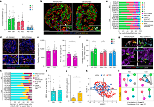
(a) High-throughput traditional immunohistochemistry (IHC) with whole-slide imaging was applied across pancreas head, body, and tail regions for the entire donor cohort, and in parallel, a subset of samples was analyzed with a 28-marker panel using co-detection by indexing (CODEX), a multiplexed technique enabling fluorescence-based imaging of large tissue sections without tissue destruction to spatially resolve many cellular phenotypes. A full list of inclusionary and exclusionary markers for cellular phenotypes analyzed is provided in Supplementary Table 4. (b-g) Analysis by traditional IHC of tissue from n = 11 ND and n = 20 T2D independent donors. (b) Pancreas weight measured during organ procurement was used to calculate endocrine cell mass in Fig. 2a. (c-g) Cross-sectional area (c-d) and cytonuclear quantification (e-g) of β cells (CPEP; green), α cells (GCG; red), and δ cells (SST; blue). Individual donor data shown in stacked bar graphs (c, e); stratification by pancreas region (d, g) includes horizontal lines (solid, ND; dotted, T2D) for mean values from combined analysis (‘Aggregate’). Donor-to-donor variability in β and α cell ratio was notable, underscoring the challenge in working with heterogeneous human tissues113, but no differences were detected between groups in this cohort30,114,115,116. (h-j) Analysis by CODEX of tissue from n = 6 ND and n = 10 T2D independent donors. (h) Cross-sectional area of endocrine cell types as measured by CODEX, including rarer γ and ε cell populations. (i) Correlation of paired cell type measurements from traditional IHC (x-axis) and CODEX (y-axis); the two methods were highly concordant (R2 = 0.8499, slope=0.9828, P < 0.0001; two-tailed linear regression). (j) Abundance of endocrine and non-endocrine cells in ND and T2D islets; one vertical bar per islet and colored by cell type. Islets are grouped by donor and ordered from largest (highest total cell number) to smallest. See also Fig. 2c. (k) Rare cells positive for chromogranin A (CHGA; red) but negative for all hormones (green), postulated to represent dedifferentiated endocrine cells117, were present in both ND and T2D at similar proportions. Scale bars, 50 μm; arrowheads denote CHGA+ hormone– cells. (l) Amyloid prevalence (% total islets with amyloid, averaged over multiple regions) for n = 11 ND and n = 20 T2D independent donors. (m) Correlation of amyloid prevalence with β, α, and δ cell populations as percentage of total endocrine cell number or cross-sectional area; one symbol per donor with 95% confidence interval of linear regression (shading). No slopes were significantly nonzero at P < 0.01 threshold. Traditional IHC data: panels c-h, l-m; CODEX data: panels i-k. Data in panels b, d, f-h, and l show mean + SEM; statistical results (b, l: two-tailed t-test; d, f-h: two-way ANOVA with Šídák’s multiple comparisons test) indicated as follows: *P < 0.05, **P < 0.01, ***P < 0.001, ****P < 0.0001. None of the variables shown had a statistically significant association with disease duration (Pearson correlation, threshold P < 0.05).
Extended Data Fig. 4 (related to Fig. 2). Integration of multiplexed imaging and transcriptional profiling highlight disrupted capillaries and immune cells within T2D islets.
(a) Gene expression fold-change (DESeq264; two-sided and multiple hypothesis corrected at FDR < 0.05) of selected vascular and neuronal ligands and their receptors in β cells, α cells, and islets; • FDR < 0.05; * FDR < 0.01. Alpha cells expressed more angiogenic ligands and receptors than β cells. (b-c) RNA-sequencing analysis highlighted enrichment in T2D samples for processes controlling blood vessel size, particularly in α cells, as well as regulation of growth factors critical to islet capillary maintenance. Panel (b) shows select vascular-related ontology terms (RNA-Enrich66; two-sided and multiple hypothesis corrected at FDR < 0.05); panel (c) shows Metascape visualization of select terms enriched for differentially expressed genes (two-tailed hypergeometric test)69 in T2D α cells (left) and islets (right). (d) Average distance of each endocrine cell type to nearest capillary (n = 6 ND and n = 9 T2D independent donors). Interestingly, α and δ cells were slightly closer to capillaries than β cells in both ND and T2D islets. (e) Phenotypes of endothelial cells (CD31; red) defined by single or dual positivity for HLA-DR (green) and CD34 (blue). Examples of each combination (HLA-DR+ CD34–, CD34+ HLA-DR–, HLA-DR+ CD34+, and HLA-DR– CD34–) are shown to right. Data from n = 6 ND and n = 10 T2D independent donors. Scale bars, 25 μm. (f-g) Enrichment in T2D β cells and islets for cytokine signaling and immune cell recruitment pathways118,119. Panel (f) shows select immune-related ontology terms (RNA-Enrich66; two-sided and multiple hypothesis corrected at FDR < 0.05); panel (g) shows magnification of select clusters depicted in Fig. 1j (terms enriched across β, α, and islet samples). (h-i) Macrophages (IBA1+) and T cells (CD3+) phenotyped by various cell surface markers; insets show additional cells to illustrate phenotypic variety. Proinflammatory (HLA-DR+), anti-inflammatory (CD163 and/or CD206+), helper (CD4+), and cytotoxic (CD8+) phenotypes are detailed in Supplementary Table 4. Scale bars, 25 μm (h, i inset) and 50 μm (i). Data from n = 6 ND and n = 10 T2D (h) or n = 5 ND and n = 8 T2D (i-j) independent donors. (j) Expression of HLA-DR in CD4+ and CD8+ T cell populations. (k) High-dimensional component analysis of islet cell composition per islet (n = 681), shown by donor; corresponds to Fig. 2k. RNA data: panels a-c, f-g; * FDR < 0.05; CODEX data: panels d, h-j. Bar graphs in panels d-e and h-j show mean + SEM with symbols representing individual donors; statistical results (d: one-way ANOVA with Tukey’s multiple comparisons test; e, h-i: two-way ANOVA with Šídák’s multiple comparisons test; j: two-tailed t-test) indicated as follows: * P < 0.05, ** P < 0.01, *** P < 0.001, **** P < 0.0001. None of the variables shown had a statistically significant association with disease duration (Pearson correlation, threshold P < 0.05).
Extended Data Fig. 5 (related to Fig. 2). Cellular neighborhood assignment and (d-e) corresponding cell composition changes in T2D and ND islets.
(a) Heat map showing cellular neighborhoods (CNs) identified by CF-IDF method and representative islet image with cell and CN annotation overlay. See Methods for more details. Scale bar, 50 μm. (b-c) A k-means cellular neighborhood analysis (b) was applied in parallel, and results were highly concordant between the two methods (c; R2 = 0.9542, slope = 1.058, P < 0.0001; two-tailed linear regression). Scale bar (b), 50 μm. Bar graphs show mean + SEM with symbols representing individual donors. (d-e) Differential cell type enrichment (two-tailed t-test) in T2D vs. ND islets by CF-IDF (d) and k-means (e) analyses. ECs and pericytes were depleted in β CNs (CN1) of T2D islets, consistent with our findings of decreased proximity between β cells and ECs in T2D. Symbols indicate unadjusted P-values as follows: ◦ P < 0.2, • P < 0.1, *P < 0.05. (f-g) Correlation of cell compositions across CNs; these analyses ask whether cell type frequencies are correlated between CNs, i.e., if there was evidence for connectivity between spatially distinct regions. Panel (g) shows results from k-means method; see also Fig. 2l (CF-IDF method). All cellular neighborhood data is derived from CODEX imaging of tissue from n = 6 ND and n = 10 T2D independent donors.
Extended Data Fig. 6 (related to Fig. 3). WGCNA emphasizes α and islet cell gene modules associated with donor and islet traits as well as those enriched in GWAS loci.
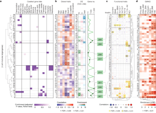
(a-d) Though α cell modules showed weaker correlations to donor and functional traits than did β cell modules, several modules were significantly enriched for cilia-related genes (a) and α08 was also enriched for α cell genes differentially expressed in T2D α cells (b). Both α08 and α16 significantly inversely correlated with epinephrine-mediated glucagon secretion and were closely related across functional parameters (c). Module α08 showed significant enrichment for T2D GWAS variants (d). See also Supplementary Fig. 4b. (e-g) Several islet modules showed notable enrichment for immune- and matrisome-related genes (e). Module i25 correlated positively with T2D status (f) and inversely with basal insulin secretion and GSIS, while i26 correlated inversely with KCl-mediated insulin secretion (g). See also Supplementary Fig. 4c. Panels a and e show module eigengenes clustered by similarity and relative enrichment of curated gene lists (see also Supplementary Table 5). Panels b and f provide correlation to donor characteristics, enrichment of differentially expressed (DE) genes, and total number of genes per module (• P < 0.05; * P < 0.01). Modules of interest highlighted (b: red, f: blue). Panels c and g show module correlation to α and β cell function (Fig. 1); significant associations highlighted (yellow). For islets (g), modules were correlated to both insulin and glucagon secretion (G + IBMX, 16.7 mM glucose with 100 μM isobutylmethylxanthine; 16.7 G, 16.7 mM glucose; 16.7 G 1°, first phase; 16.7 G 2°, second phase; 1.7 G+Epi, 1.7 mM glucose and 1 μM epinephrine; KCl, 20 mM potassium chloride). Panel d shows α cell module enrichment for GWAS traits (FIns, fasting insulin; 2hGlu, 2-hour glucose; FGlu, fasting glucose; * FDR < 0.01). Panels a, e, b and f (DE genes): two-tailed Fisher test adjusted for multiple comparisons; panels c, g, b and f (donor traits): t-test, unadjusted, using Spearman correlations (see Methods for additional details); panel d: enrichment using GARFIELD37.
Extended Data Fig. 7 (related to Fig. 3). Both transcriptomic and histologic data suggest changes in cilia processes in early T2D.
(a) Magnification of select clusters depicted in Fig. 1j (terms enriched across β, α, and islet samples). (b) Fold change of validated cilia-related genes (shown here, those ≥ |1.5| in both α and β cells); the majority were expressed at higher levels in T2D compared to ND. (c-d) Visualization by immunohistochemistry of cilia (ARL13B; red) and quantification of abundance, density, and size in tissue from n = 8 ND and n = 8 T2D independent donors. Tissue sections are from the same donors shown in panel b. Scale bars, 50 μm. Bar graphs show mean + SEM with symbols representing individual donors; statistical results (d: two-tailed t-test) indicated as follows: * P < 0.05, ** P < 0.01, *** P < 0.001, **** P < 0.0001.
Extended Data Fig. 8 (related to Fig. 4). RFX motif enrichment across β cell modules and β cell RFX6 reduction in pseudoislets impairs insulin secretion.
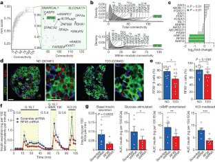
(a) Enrichment of selected transcription factor120 motifs in β cell modules, calculated using the AME tool from MEME-Suite with Bonferroni correction77; log10(P-values) are unadjusted. (b) Schematic of adenoviral shRNA delivery and formation of pseudoislets, which mirror native human islet architecture26,121. (c) Relative RFX6 mRNA expression in β cells treated with scramble shRNA (‘control’) or RFX6 shRNA (‘shRFX6’). Data represents n = 3 independent islet preparations. (d) RFX6 knockdown did not change β or α cell proportion (n = 4 independent islet preparations); acute (6-day) reduction of RFX6 expression does not lead to β cell loss. (e) Control and shRFX6 pseudoislets exhibited similar size and morphology. Transduced cells marked by mCherry; distribution of β cells shown by C-peptide (CPEP; blue) and α cells by glucagon (GCG; green). Scale bars, 200 μm (morphology) and 50 μm (immunostaining). (f-i) Dynamic insulin secretion and metrics equivalent to Fig. 4f, g but normalized by total insulin content (h) in control and shRFX6 pseudoislets. (i) Proinsulin content and proinsulin:insulin ratio in control (scramble) and shRFX6 pseudoislets. Proinsulin content was not significantly greater in shRFX6 pseudoislets and the proinsulin:insulin ratio was comparable to controls, suggesting secretory defects were not due to inappropriate insulin processing. Functional data (f-i) represents n = 6 independent islet preparations. Data in panels c-d and f-i show mean + SEM; statistical results (c-d, g-i: two-tailed t-test; f: linear mixed-effect model) indicated as follows: *P < 0.05, **P < 0.01, ***P < 0.001, ****P < 0.0001.
Extended Data Fig. 9 (related to Fig. 5). RFX6 controls stimulated insulin secretion through genome-wide chromatin alterations disrupting transcripts controlling exocytotic pathways.
(a) Post-QC nuclei counts from control and shRFX6 pseudoislets. (b) Abundance of fluorescent marker gene expression (mCherry/mKate2) in α and β cell nuclei. Reporter expression was much higher in β cell nuclei than in α cell nuclei, consistent with the previously observed preferential adenoviral targeting of β relative to α cells26. (c) Proportion of differentially expressed (DE) genes per cell type. Nuclear RFX6 was not among those reduced in β cell nuclei, consistent with shRNA silencing occurring in the cytoplasm. (d) Pathway enrichment (g:Profiler75, two-tailed hypergeometric test) for DE genes (FDR < 0.01); second two columns separate genes up- or downregulated in shRFX6. (e-f) To identify gene regulatory networks driving cell states, we employed Single-Cell rEgulatory Network Inference and Clustering (SCENIC) and discovered cell type specific regulons governing cell identity. (f) UMAP clustering of nuclei (RNA component) by SCENIC using regulon activity (left) and average regulon activity across cell types (right). See also Fig. 5d. (g) To investigate overlap in differentially expressed genes between shRFX6 β cell nuclei and sorted T2D β cells, we compared the top 1,000 most significant DE genes in shRFX6 vs. control β cell nuclei (blue) and T2D vs. ND sorted β cells (red). Circos plot illustrates commonality at the level of gene IDs (purple) or ontology term enrichment (grey; p < 0.01). Metascape network analsyis69 displays a subset of enriched terms, where edges denote term similarity and node colors represent contribution of each gene list. Common pathway enrichment related to microtubule cytoskeleton organization, ion transport, and regulation of protein secretion. (h-i) Motif enrichment for top 2,000 (h) or 10,000 (i) RFX6-sensitive up- and downregulated ATAC peaks in shRFX6 β cell nuclei. Motifs with highest significance are labeled in top panels; significant RFX motifs (or the single RFX motif closest to significance, in the case that no RFX motifs reach significance) are labeled in bottom panels. (j) Enrichment of top RFX6-sensitive up- and downregulated ATAC peaks (n = 2,000, 5,000, or 10,000) in shRFX6 β cell nuclei near shRFX6 β cell differentially expressed genes. (k-l) Single value odds ratio of T2D GWAS enrichment (k) and model estimate from conditional analysis (l) of top 2,000 or 10,000 RFX6-sensitive peaks; see Methods for details. Nominal P-values for panel k, left to right: 0.00336, 5.53e-28, 0.0194, 9.28e-22. (m) Model of Mendelian randomization (MR) depicting the relationship of an instrumental variable (here, RFX6 SNPs) related to exposure (RFX6 expression) and outcome (T2D). Dotted lines denote assumptions of no association while solid and dashed lines indicate disease causality. (n) MR analyses using the European ancestry cohort from Vujkovic et al. meta-analysis9 (n = 1,114,458 independent samples) resulted in point estimates that were directionally consistent with the findings for the UK Biobank (see Fig. 5k). The heterogeneity across contributing studies (for example Million Veterans Program data reflect an older T2D case set, more comorbidities, and a strong male bias compared to UK Biobank) likely reduces our power. We leveraged several MR approaches to determine whether results were robust to their varying assumptions. Debiased inverse variant weighted method (robust to weak instrument bias): causal effect = −0.084, P = 0.065; Pleiotropy RESidual Sum and Outlier (PRESSO) method (removes outlier instrumental variable effects): causal effect = −0.073, P = 0.251; weighted median method (robust when up to 50% of instrumental variables are invalid): causal effect = −0.045, P = 0.372. Results from MR-Egger are not shown due to strong weak instrument bias (I2_GX = 0). Error bars in panels l and n represent 95% confidence intervals.
Extended Data Fig. 10 (related to Fig. 5). Summary schematic of key molecular alterations in short-duration T2D cohort and of the convergence of genetic risk on an RFX6-mediated gene regulatory network.
(a) Major β cell-intrinsic and islet microenvironment alterations that define islet dysfunction in early T2D. Observations from transcriptomic and histologic studies revealed no change to endocrine cell composition but evidence of dysregulated β cell processes and modest changes to intraislet vascular and immune cell populations. Insulin secretion was reduced and persisted in a nondiabetic environment; glucagon hypersecretion was not observed122. (b) After identifying RFX6 as a candidate disease-associated gene through unbiased analysis of a small cohort (left panel; labels 1-2), we used two approaches for validation: first, molecular perturbation of RFX6 in β cells of primary human pseudoislets allowed functional, transcriptomic and epigenomic analyses (top right panel; labels 3-4) and second, integration of UK Biobank data allowed population-scale genetic relationship to be examined (bottom right panel; label 5). Reduction of RFX6 led to reduced insulin secretion defined by transcriptional dysregulation of vesicle trafficking, exocytosis, and ion transport pathways that was mediated by genome-wide chromatin architectural changes overlapping with T2D GWAS variants. Furthermore, Mendelian randomization analysis revealed that reduced islet RFX6 expression is causally associated with T2D.
Supplementary information
Supplementary Figures
Supplementary Figs. 1–6.
Supplementary Tables
Supplementary Tables 1–6.
Supplementary Data 1
This file contains: Supplementary Data 1a: genes that are differentially expressed at Padj < 0.01, T2D vs ND, bulk RNAseq; Supplementary Data 1b: pathway enrichment (Metascape) of differentially expressed genes at Padj < 0.01, T2D vs ND, bulk RNAseq; and Supplementary Data 1c: GOBP pathway enrichment (RNA-enrich) of differentially expressed genes at Padj < 0.01, T2D vs ND, bulk RNAseq.
Supplementary Data 2
This file contains: Supplementary Data 2a: genes that are differentially expressed at Padj < 0.01, shRFX6 vs scramble, β cell nuclei, multiome (RNA); Supplementary Data 2b: pathway enrichment (Metascape) of differentially expressed genes at Padj < 0.01, shRFX6 vs scramble, β cell nuclei, multiome (RNA); Supplementary Data 2c: genes connected to top 10 differential β regulons, shRFX6 vs scramble, β cell nuclei, multiome (RNA); Supplementary Data 2d: pathway enrichment (Metascape) of genes connected to top 10 differential β regulons, shRFX6 vs scramble, β cell nuclei, multiome (RNA); Supplementary Data 2e: pathway enrichment (Metascape) of differentially expressed genes at Padj<0.01; shRFX6 vs scramble, β cell nuclei, multiome (RNA) and T2D vs ND, bulk RNAseq.
Supplementary Data 3
Effect estimates for genetic association with RFX6 expression and T2D.
Supplementary Information
The HPAP Consortium.
Rights and permissions
Springer Nature or its licensor (e.g. a society or other partner) holds exclusive rights to this article under a publishing agreement with the author(s) or other rightsholder(s); author self-archiving of the accepted manuscript version of this article is solely governed by the terms of such publishing agreement and applicable law.
About this article
Cite this article
Walker, J.T., Saunders, D.C., Rai, V. et al. Genetic risk converges on regulatory networks mediating early type 2 diabetes. Nature (2023). https://ift.tt/JXWCGTE
-
Received:
-
Accepted:
-
Published:
-
DOI: https://ift.tt/JXWCGTE
Comments
By submitting a comment you agree to abide by our Terms and Community Guidelines. If you find something abusive or that does not comply with our terms or guidelines please flag it as inappropriate.
"type" - Google News
December 04, 2023 at 11:31PM
https://ift.tt/6zPVOx2
Genetic risk converges on regulatory networks mediating early type 2 diabetes - Nature.com
"type" - Google News
https://ift.tt/HP0Garu
https://ift.tt/6Z5JSlu
Bagikan Berita Ini


0 Response to "Genetic risk converges on regulatory networks mediating early type 2 diabetes - Nature.com"
Post a Comment